Scientists invent a camera that can measure the size and location of tumors
A camera that could help surgeons locate and remove tumors has been developed by two US scientists

The best camera deals, reviews, product advice, and unmissable photography news, direct to your inbox!
You are now subscribed
Your newsletter sign-up was successful
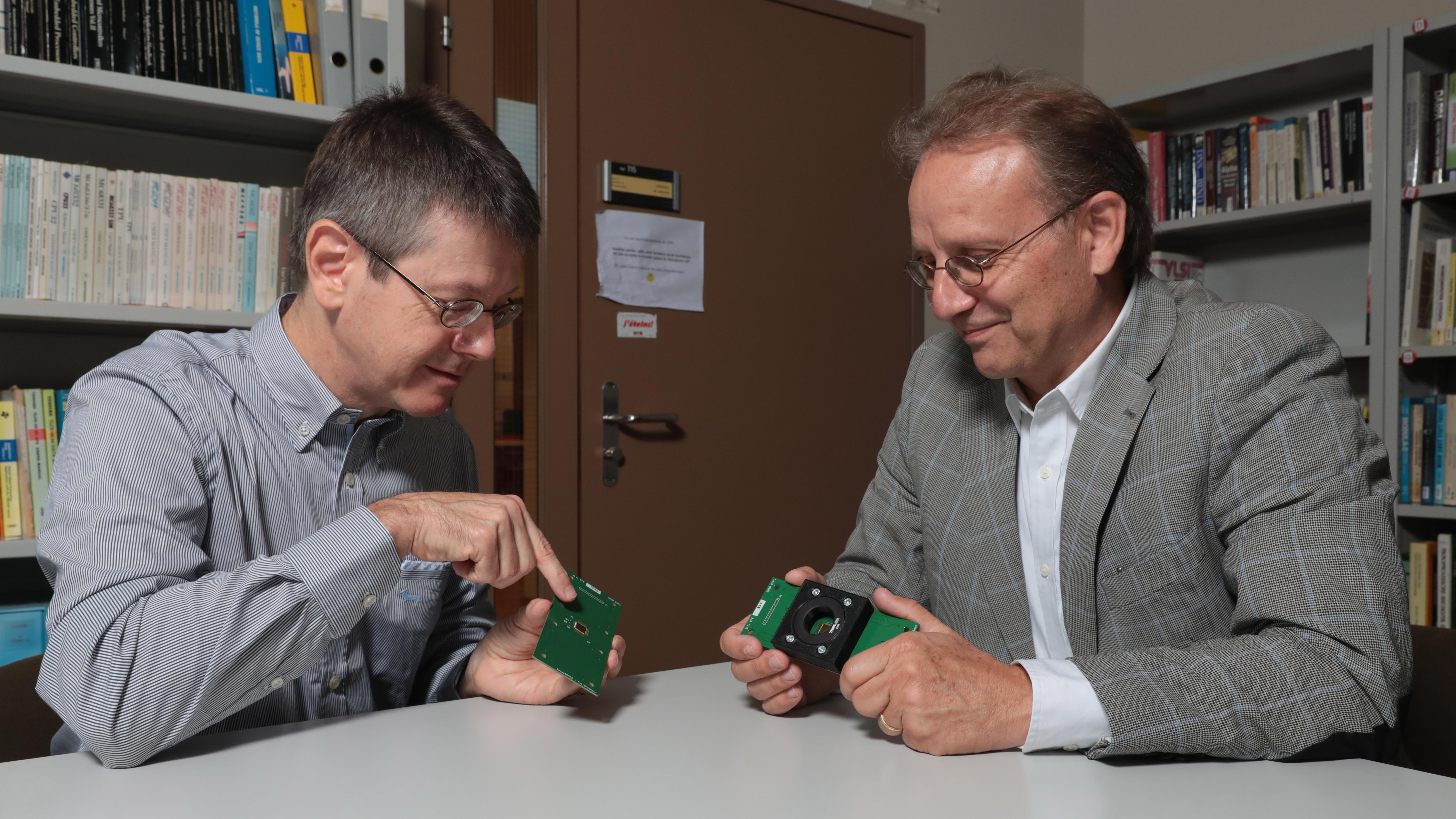
A camera capable of detecting the exact size and position of tumors has been developed by two scientists in the US. Edoardo Chabon of EPFL (École polytechnique fédérale de Lausanne) and Claudio Bruschini of Dartmouth College worked together in the hope the camera will help surgeons remove cancerous cells.
Edoardo Chabon, head of the Advanced Quantum Architecture Laboratory at EPFL, invented the camera known as the SwissSPAD2. It was able to capture and count the very smallest form of light particles known as photons, while also being able to generate 3D images and calculate the depth of field. It works by measuring the amount of time it takes for a photon to travel from the camera to an object using a photon-counting pixel sensor and a pulsating laser.
• Read more: Best microscopes
Working alongside Claudio Bruschino to push the technology further, the two have come up with a method that involved projecting red light onto an area of diseased tissue with a lazer while simultaneously taking a picture of the area. “Red is a color that can penetrate deep into human tissue,” Chabon explained to Mirage News, and when these red light particles reach a tumor they behave differently.
The tissue being examined is injected with a fluorescent contrasting agent that attaches only to the tumor cells, and this helps identify how long it takes for the light particles to return to the point they came from. The time difference between when the red light particles return in healthy tissue and tumorous tissue is what is needed to reconstruct the tumor.
There is less than a nanosecond delay in the time it takes for the particles to return, but that is enough for the scientists to be able to generate 2D and 3D images. It’s this attribute that enables the system to identify a tumor's shape, thickness and location in a patient's body.
Although surgeons can use MRI scans to locate tumors in the body post-op, it’s a lot harder to locate them from healthy tissue during surgery. This new technology is designed to help surgeons complete the delicate task of removing a tumor and ensure that no cancerous tissue is left behind – and hopefully help to save lives.
The best camera deals, reviews, product advice, and unmissable photography news, direct to your inbox!
Read more:
Scientists invent a camera so small a beetle can carry it
Best thermal imaging cameras
How to build a microscope with a smartphone and lego

Having studied Journalism and Public Relations at the University of the West of England Hannah developed a love for photography through a module on photojournalism. She specializes in Portrait, Fashion and lifestyle photography but has more recently branched out in the world of stylized product photography. Hannah spent three years working at Wex Photo Video as a Senior Sales Assistant, using her experience and knowledge of cameras to help people buy the equipment that is right for them. With eight years experience working with studio lighting, Hannah has run many successful workshops teaching people how to use different lighting setups.
